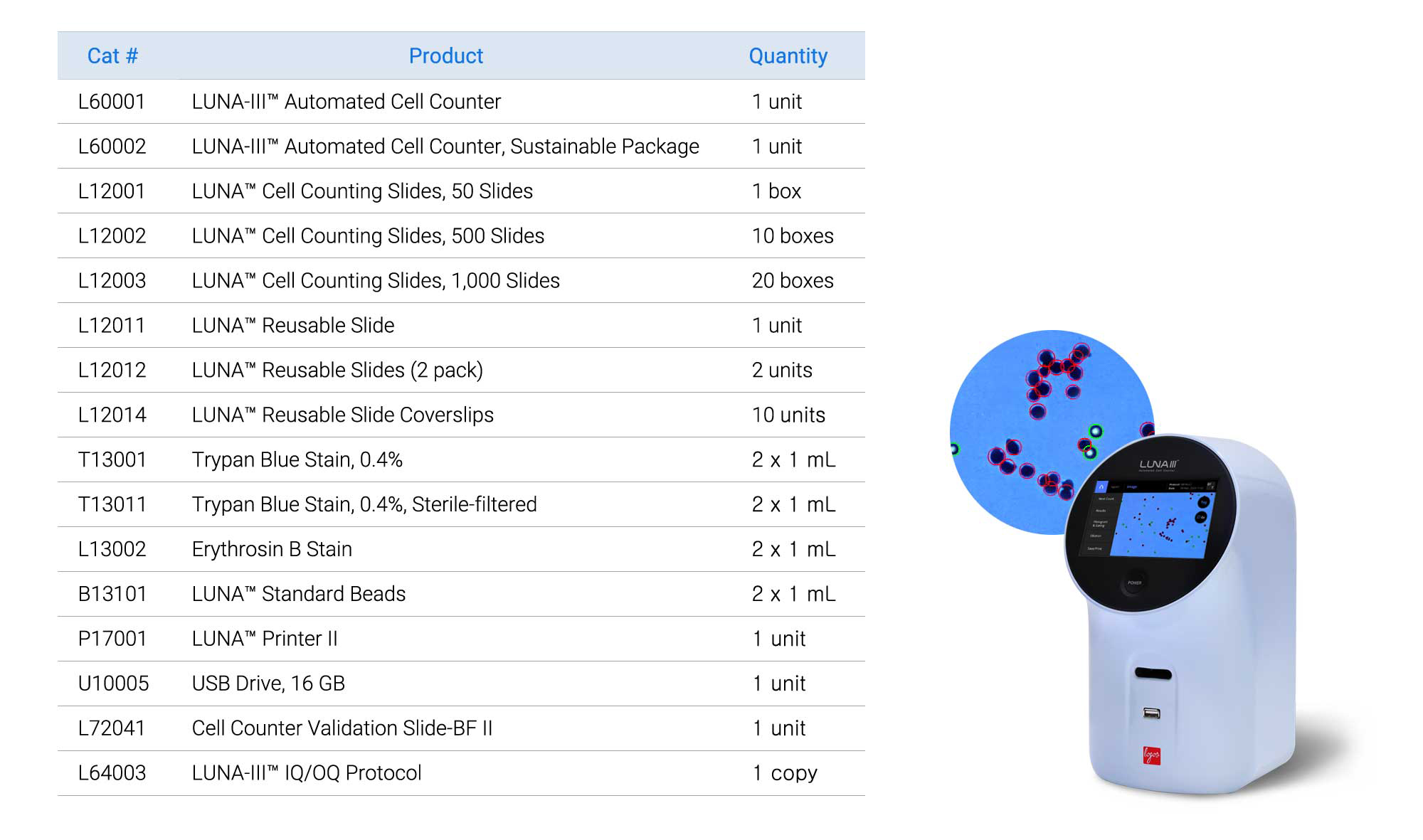
Products
Customer Reviews
Resources
Support
Keywords: LUNA-III™ Automated Cell Counter, PBMC, offset feature, trypan blue staining, cell concentration and viability assessment
Peripheral Blood Mononuclear Cells (PBMCs) play a crucial role in research areas, including infectious diseases, autoimmune disorders, and cancer immunology. Consistency and reproducibility in these studies are essential for reliable and accurate outcomes. However, accurately counting PBMCs can be challenging due to both their characteristics and sample quality. Their small size and dim edges can compromise detection accuracy. Additionally, PBMC preparations often contain a high level of debris and potential red blood cell (RBC) contamination, as they are derived from blood. These factors can negatively affect autofocusing performance, leading to inaccurate cell counting and viability measurements.
Given these limitations, fluorescence-based cell counters are generally more suitable, and products like the LUNA-FX7™ automated cell counter are often recommended for such purposes. Achieving accurate PBMC counts in an affordable setting with only a brightfield optical system can be challenging. To address this, a new strategy was developed for the LUNA-III™ automated cell counter, incorporating a focal plane offset and an enhanced cell detection algorithm. Staining solutions like trypan blue (TB) can further enhance detection accuracy by making cell edges more visible. This approach aims to improve the accuracy and precision of PBMC counting and viability assessments, providing a reliable yet affordable solution. In this application note, we validate the effectiveness of the LUNA-III™ automated cell counter by comparing its PBMC data with the renowned LUNA-FX7™ automated cell counter.
Human peripheral blood was processed to obtain a PBMC sample using the standard density gradient centrifugation technique with Histopaque-1083 (Sigma, #10831). After the final wash, PBMCs enriched in the buffy coat were resuspended in PBS containing 1% BSA.
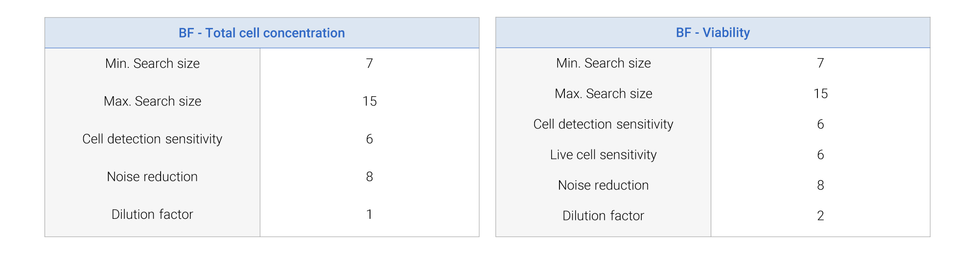
For serial dilution measurements, the cells were diluted with different dilution factors from 1 to 32. A separate batch of cells was prepared for viability measurements with theoretical viability level of 0, 25, 50, 75, and 100%. Cell counts and viability measurements were performed on the LUNA-III™ using the LUNA™ Cell Counting Slide and the built-in PBMC protocol (Table 1). Before loading, the cells were mixed with 0.4% TB (Cat No.T13001) in a 1:1 ratio. The same samples were also used in LUNA-FX7™ to compare the results.
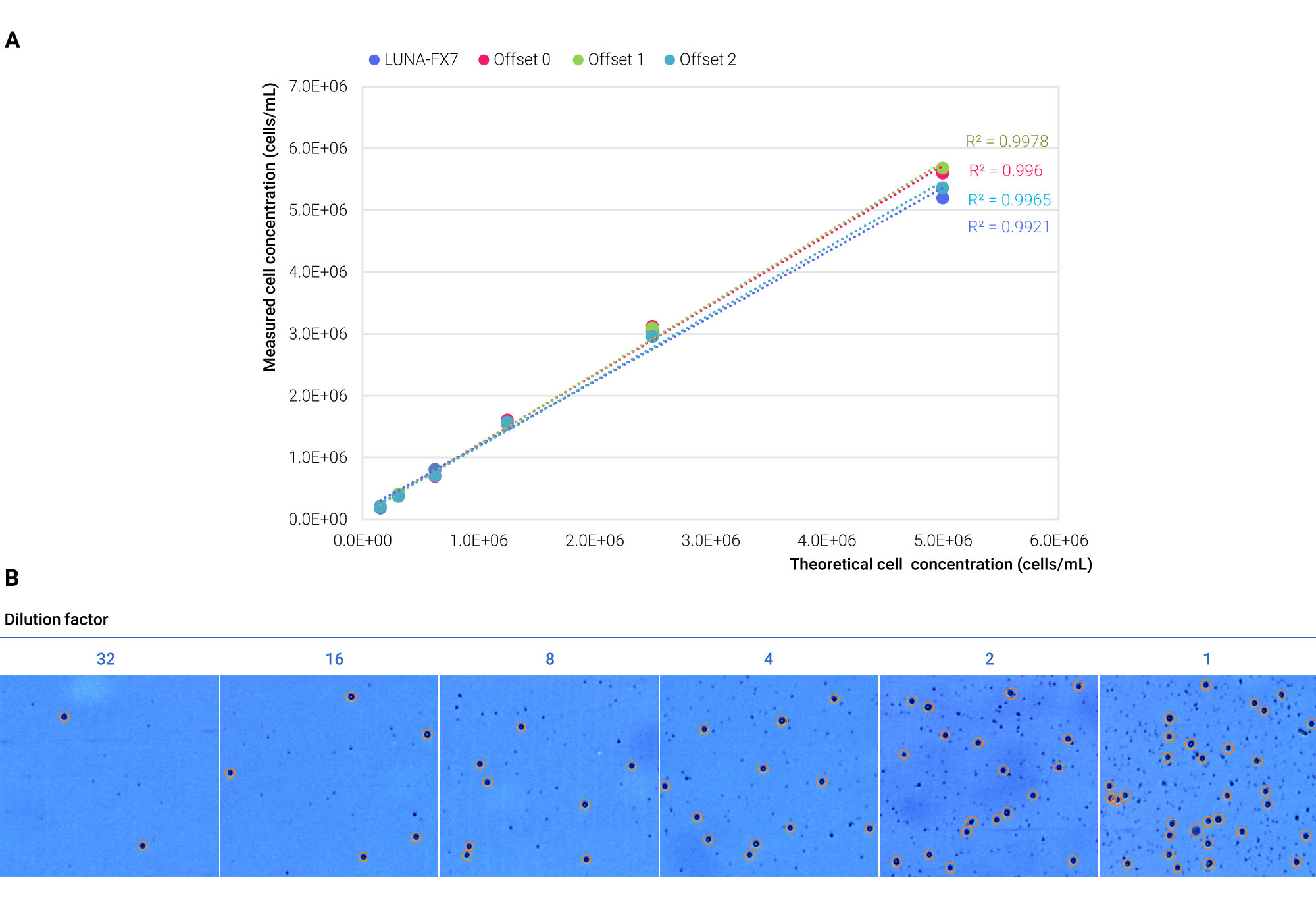
Assessing PBMC Concentration and Viability Using Different Offset Levels on the LUNA-III™
PBMC samples were tested using three offset levels (0, 1, and 2) the LUNA-III™ and LUNA-FX7™ to confirm linearity across various concentration and viability levels.
Cell concentrations at all offset levels showed a strong correlation, with an R² value of 0.9921 or higher (Figure 1A). Despite the strong linearity across all offset levels, offset level 2 yielded results closest to those obtained using the LUNA-FX7™. Moreover, PBMCs were accurately tagged using the LUNA-III™ (Figure 1B).
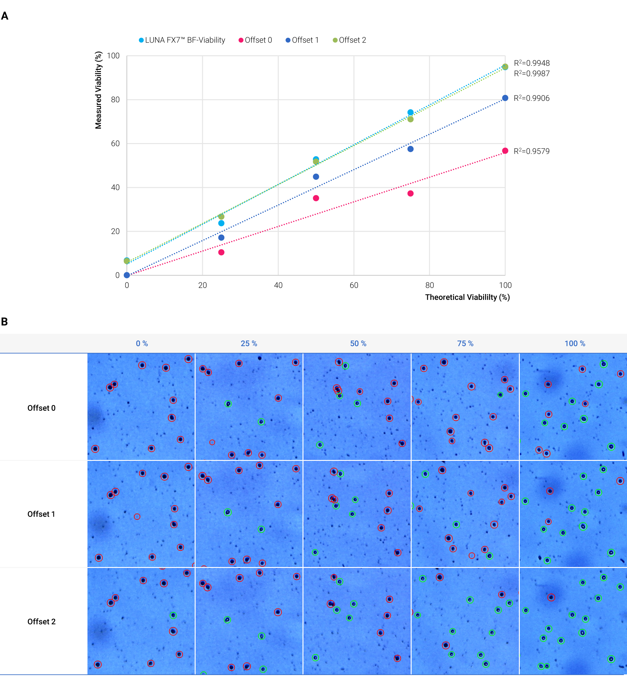
Viability measurements were conducted at three offset levels (0, 1, and 2) alongside LUNA-FX7™ viability data. While offset levels 1 and 2 showed strong correlations with theoretical viability (R² = 0.9906 and 0.9987), only the values from offset 2 aligned with those from LUNA-FX7™ (Figure 2A). Offset level 0 showed a lower correlation (R² = 0.9579) with theoretical values, with its results deviating considerably from LUNA-FX7™. Live and dead cell tagging was most accurate at offset level 2 across different viability levels when using the LUNA-III™ system (Figure 2B).
Limitations of Using Trypan Blue Staining for PBMC Counting
Despite our results demonstrating the effectiveness of TB staining for PBMCs using the LUNA-III™, this method has notable limitations for PBMC counting. One major issue with TB staining is its inability to differentiate between PBMCs and contaminants, such as red blood cells (RBCs) and debris. This can lead to overestimation of total cell counts and an underestimation of cell viability, as contaminants can be erroneously counted as dead cells. In contrast, fluorescence (FL) staining methods, such as those used in advanced fluorescence-based cell counters like the LUNA-FX7™, provide a more accurate assessment by selectively identifying live and dead cells while effectively excluding contaminants. Thus, for PBMC samples that may contain RBCs or debris, fluorescence staining is strongly recommended to ensure precise results, particularly when high accuracy in viability and total cell counts is critical.
The integration of a focal plane offset and advanced cell detection algorithm in the LUNA-III™ has significantly improved the accuracy and precision of PBMC counting and viability assessments, with offset level 2 closely matching the high performance of the LUNA-FX7™. However, it is essential to recognize that while the LUNA-III™ offers a cost-effective solution, using trypan blue (TB) staining can present challenges. TB staining is unable to differentiate PBMCs from contaminants like red blood cells and debris, potentially leading to overestimated cell counts and inaccurate viability readings, as contaminants may be miscounted as dead cells. Therefore, users must consider these limitations when working with PBMC samples, especially when higher accuracy is required in viability measurements.